Electromyography (EMG). Everything you should know
Electromyography (abbreviated EMG) is a diagnostic procedure for examining muscles and the nerves (motor neurons) that supply them. EMG results can reveal dysfunctions of nerves and muscles or problems with nerve-to-muscle signal transmission.
You've probably had an electrocardiogram (ECG/EKG) before. During that test the device recorded the electrical activity of your heart. Well… an electromyogram (EMG) is made for a similar purpose, but it examines your skeletal muscles and their electrical activity rather than the heart muscle.
The electrical impulses that control your movements travel from your central nervous system to your muscles via motor nerves, causing muscle contraction. During an electromyography exam, the electrical activity that occurs during this process is recorded.
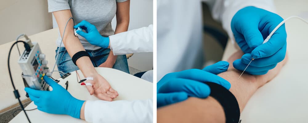
The test can be performed using electrodes attached to the skin surface or with needle electrodes inserted into the muscle. In surface testing, they examine the magnitude and speed of electrical activity between two or more points — that is, the signal conduction ability of the motor nerve. Needle electrodes inserted into the muscle record the response generated within the muscle.
Why is an EMG performed?
An EMG is performed when you have symptoms suggesting a nerve or muscle disorder. These may include, among others:
- Tingling
- Numbness
- Muscle weakness
- Muscle pain or recurrent cramps
- Certain types of limb pain
- Paralysis
- Muscle wasting
- Muscle twitching
Below I list some conditions and diseases related to muscles and motor nerves. Analyzing electromyography results can help establish a diagnosis or (just as importantly) rule out a condition.
Muscle disorders
- For example, muscular dystrophy, which most often begins in childhood and primarily affects boys. Some types develop only in adulthood. All forms of the disease are genetically determined, meaning their development is always linked to genetic defects. If muscular dystrophy affects the neck and chest muscles, breathing and swallowing may also become difficult. Contractures can develop due to the opposing muscles (antagonists) acting against the wasting muscles, which can pull limbs into an "out of position" or twisted posture. It is not curable, though some interventions can slow the progression.
- Polymyositis is a rare myopathy that leads to inflammation and muscle weakness. It causes discomfort and even pain in the muscles. It generally affects people between 30 and 60 years of age and is more common in women than in men. The immune system attacks muscle fibers, causing damage and inflammation. Some experts think a virus may trigger the condition or that it may appear as an allergic reaction after certain medications. Some genes may also influence the risk of polymyositis. There is no cure, but there are therapies that help control inflammation and relieve symptoms.
Disorders affecting the connection between nerve and muscle
- Myasthenia gravis. A severe muscle weakness and abnormal fatigability of muscles due to autoimmune causes. The transmission of nerve impulses between nerves and muscles is impaired. It can affect both women and men, but is more common in women. It can occur at any age, but typically appears between 20 and 40 years. The trigger of the autoimmune process is unknown, but it has been observed that affected individuals often have a thymus gland that has not regressed as expected. Patients usually present to their doctor with drooping eyelids (ptosis) or double vision. The disease is not currently curable, but symptoms can be kept under control with therapy.
Peripheral nerve disorders
Various disorders can affect the nerves outside the spinal cord (that is, peripheral nerves).
- Carpal tunnel syndrome (or wrist tunnel syndrome). It is caused by inflammation around the tendons that flex the fingers, so treatment focuses on effective anti-inflammatory measures. Nowadays more people work on computers, which has led to various joint/musculoskeletal problems. In carpal tunnel syndrome, increased pressure is placed on the median nerve within the C-shaped tunnel at the wrist formed by tendons and bones. Since some of the tendons and nerves that run from the hand to the forearm pass through the carpal tunnel, disturbances there cause nighttime numbness and pain in the fingers and, in severe cases, wasting of the thumb pad muscles.
- Neuropathy. Refers to diseases of the nerves outside the brain and spinal cord (that is, peripheral nerves). In developed countries it most often develops as a complication of diabetes. It can also be associated with autoimmune diseases such as rheumatoid arthritis or systemic lupus erythematosus (SLE), certain vitamin deficiency states such as vitamin B1 deficiency, chronic alcoholism, and some medications. Treating the underlying disease is often sufficient; other times peripheral neuropathy can be alleviated by managing the pain.
Diseases affecting motor neurons in the brain and spinal cord
- Amyotrophic lateral sclerosis (ALS). A fatal disease characterized by the degeneration of motor nerve cells in the brain and spinal cord that innervate voluntary muscles. The incidence is about 5 patients per 100,000 population, which for Hungary's 10 million population would mean roughly 500 new cases per year. The exact cause is unknown. Motor function progressively deteriorates. Meanwhile, cognition, vision, hearing, sensation, taste, and touch remain intact. Interestingly, although voluntary muscles are involved, eye movements and bowel and bladder function often remain normal. Most patients die from respiratory failure. Progress cannot be halted; treatment aims to maintain quality of life as much as possible.
- Poliomyelitis (infantile paralysis). A syndrome caused by viruses in which motor neurons located in the spinal cord are destroyed. Due to mandatory immunizations, this infection no longer occurs in Europe today.
Conditions affecting nerve roots
- Herniated disc. A protruding disc can press on a nerve, causing sharp pain that worsens with movement. The location of the pain depends on which nerve root is compressed. A transitional sacral herniation can cause pain radiating into the legs, commonly called sciatica. Pain in the lumbar spine region usually radiates to the lower back. This is referred to as lumbago or simply back pain.
Electromyography
EMG is a low-risk procedure and complications are rare. There is a small risk of bleeding, infection, and nerve injury where the needle electrode is inserted or in the surrounding area. When muscles located along the chest wall are examined with needle electrodes, there is a very small risk that air could leak into the space between the lung and chest wall, causing lung collapse (pneumothorax).
How should you prepare for the test?
- When scheduling the EMG, ask whether you should stop taking any medications before the test. For example, if you take medication for myasthenia gravis, you should specifically ask whether you need to stop it for the examination.
- Shortly before the test, shower or bathe to remove oils from your skin. Do not apply creams or lotions before the procedure.
- Do not smoke for at least three hours before the test.
- Remove any jewelry that could interfere with the examination.
Inform the neurologist and the EMG technician if you:
- Have a pacemaker or any other implanted electrical medical device.
- Are taking blood-thinning medications.
- Have hemophilia, a bleeding disorder that causes prolonged bleeding.
What will happen?
You will need to remove clothing from at least the limb being examined. The test is usually performed with you lying on an examination table.
An assistant will place surface electrodes on different points of your skin depending on where you experience symptoms. During the test, small electrical impulses are delivered through the surface electrodes, which you may feel as a pinprick sensation on the skin or as a muscle twitch.
The neurologist may also insert needle electrodes into your muscles depending on your symptoms. The needle stick can cause discomfort or pain, which usually subsides shortly after the needle is removed. During needle EMG the neurologist examines whether there is spontaneous electrical activity when the muscle is at rest — activity that is not present in healthy muscle tissue — as well as the amount of activity when you slightly contract that muscle. The neurologist will instruct you to relax or tense the muscle. Depending on which muscles and nerves are being examined, you may need to change your body position.
If you feel uncomfortable or experience pain at any time during the test, tell the neurologist and you can take a short break.
The procedure typically takes 30–60 minutes depending on the area examined.
After the test, you may have temporary, minor bruising where the needle electrode was inserted into the muscle. This bruise will fade within a few days.
Results
The neurologist interprets the test results and prepares a summary report. You should discuss the findings and next steps with your general practitioner or the doctor who ordered the EMG.