Mammography — what you need to know
Regular participation in screening examinations could prevent a large proportion of breast cancer, which is a leading cancer among women aged 45 to 65. In addition, 'self-examination' can also play an important role in reducing the number of women who die from breast cancer. Imaging tests, such as breast ultrasound and mammography, can help early [...]

Regular participation in screening examinations could prevent a large proportion of breast cancer, which is a leading cancer among women aged 45 to 65. In addition, 'self-examination' can also play an important role in reducing the number of women who die from breast cancer. Imaging tests, such as breast ultrasound and mammography, can help early diagnosis.
'Interesting' is the fact that breast cancer is particularly common in the Western world: it occurs much more frequently in Europe and the United States than in developing and eastern countries.
Mammography — a method for examining the breast
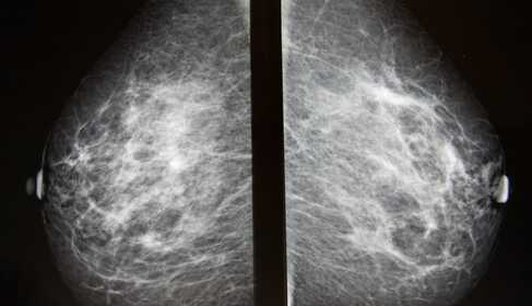
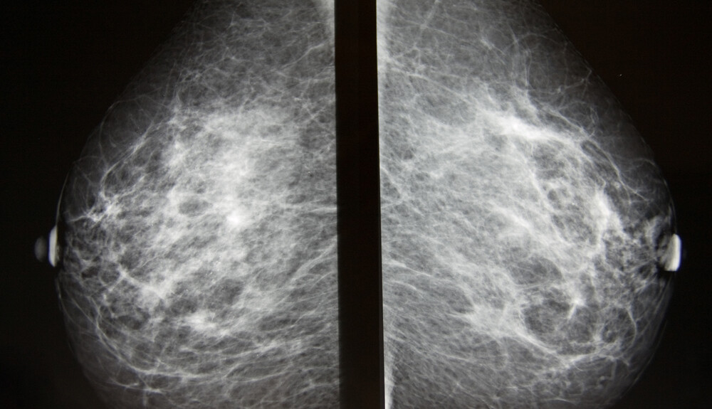
During mammography, X-ray images of the breasts are taken. These can then be used for breast cancer screening or for diagnostic purposes, for example to investigate certain symptoms or unusual findings detected by another imaging method.
During the exposure, the breast is compressed between two solid surfaces so that the breast tissue spreads out. Then black-and-white images are produced by X-ray, which are displayed on a computer screen and examined for signs of cancer. The image is taken with an X-ray machine specifically designed for mammography. To ensure good image quality and to reduce radiation exposure, each breast is compressed individually with special plastic plates and two-directional X-rays are taken of them. A conventional mammography is basically not painful, but the compression of the breasts can be uncomfortable. A technician will help position your head, arms and torso so that the machine can scan your breasts without obstruction.
Mammography plays a key role in breast cancer screening because it can detect abnormalities before they cause symptoms. It has been shown to reduce mortality from breast cancer.
With conventional imaging a two-dimensional picture of the breast is produced. A newer type, 3D mammography, produces three-dimensional images of the breast. Some healthcare facilities already offer 3D mammography alongside traditional 2D mammography for breast cancer screening.
When is it used?
The mammography exam is preceded by a detailed personal and family medical history and an assessment of possible risk factors, followed by a physical examination. After that, two-directional mammographic images of both breasts are taken, which takes approximately 5 minutes.
The X-ray machine uses low-energy ionizing radiation, so the radiation exposure is small.
Screening mammography
Screening means the examination is performed when you have no complaints and are asymptomatic. The aim is to detect cancerous changes in the breast as early as possible. If a cancerous lesion is discovered at an early stage, its treatment is simpler, less invasive and more successful.
Breast cancer can occur at quite a young age; case numbers increase in those over 35. With increasing age these numbers rise further, so the incidence of breast tumors doubles after the age of 45 and quadruples after the age of 65. Although the probability of developing breast cancer depends on many factors (breast structure, family predisposition, hormonal status), overall it is advisable to start regular annual breast screenings from the age of 40. If there has been a breast tumor in the family, breast examinations (mammography, ultrasound) may be recommended even earlier, around age 35.
Diagnostic mammography
Suspicious breast changes can be varied. For example, a lump in the breast, breast pain, unusual skin changes, thickening of the nipple or nipple discharge. In such cases, mammography is used for further investigation.
Risks
The risks and limitations of mammography include the following:
- Mammography involves a low dose of ionizing radiation. The benefits of regular mammography outweigh the risks associated with this level of radiation.
- If something 'suspicious' is seen on the mammogram, this alone is not enough — further examinations are required. These may include additional imaging studies, such as ultrasound, as well as biopsy (surgical sampling of the breast tissue containing the lesion). Histological analysis of the sample determines the further course of action.
In most cases, an abnormality seen on a mammogram is not malignant. Additional tests are necessary to determine this beyond doubt.
Screening mammography cannot detect every cancer: it may happen that something recognizable on physical examination is not visible on mammography. If it is too small, or located in an area that mammography has difficulty visualizing, such as the underarm, we cannot be completely certain of the diagnosis.
Although mammography enables early detection, not all types of cancer are curable. Some breast cancers are aggressive, grow rapidly and spread quickly to other parts of the body.
When is mammography not recommended?
- During pregnancy — in this case breast ultrasound is primarily recommended.
- If you have had a mammography within the past year — in this case breast ultrasound is primarily recommended.
How to prepare?
- The exam is recommended during the first two weeks after your period, because the breasts are less sensitive then.
- If you have had previous examinations, bring the earlier images with you. The radiologist can compare previous images with the new ones.
- Do not use deodorant before the exam. Deodorants, antiperspirants, powders, creams or perfumes may contain metal particles that can interfere with the image.
- In the case of breast implants, 3D mammography is much more effective than conventional mammography.
- The exam can be performed on breastfeeding mothers as well, but breastfeeding should be resumed only after 24 hours following the examination.
What will happen?
During the examination
Remove jewelry and necklaces. You will need to undress from the waist up, including your bra.
The examination is performed standing. You will stand in front of the X-ray machine. Your head, arms and torso will be positioned so that the machine can scan your breast without obstruction.
To ensure good image quality and to reduce radiation exposure, the breasts are compressed with special plastic plates and two-directional X-rays are taken of them.
The pressure is applied for a few seconds. This is not harmful but can cause some discomfort.
During the short X-ray exposure you must remain still and hold your breath so that there is no movement (movement would blur the image).
After the examination
After images of both breasts have been taken, the quality of the images is checked. If the images are technically inadequate, part of the examination may need to be repeated. The whole procedure usually takes less than 30 minutes. You can then dress and continue your normal daily activities.
The radiologist will evaluate the images. Based on the result, further imaging — MRI or ultrasound — may be required to establish an accurate diagnosis.
Results
As mentioned, mammography produces black-and-white images of the breast tissue. These are digital images displayed on a computer screen for the radiologist to analyze.
The radiologist looks for signs suggestive of cancer and other conditions that may require further tests, follow-up or treatment.
Ask when and how the results will be shared with you; generally they are available within a few days after the exam.