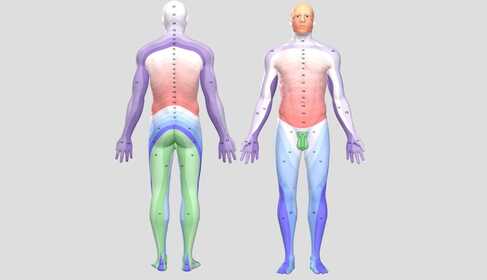

 What is a dermatome — and why is it worth knowing about even as a non-professional?
A dermatome is the strip of your skin whose sensations — touch, pain and temperature — are carried towards your brain by a single spinal nerve root. A total of 31 pairs of nerve roots leave the spinal cord (8 cervical, 12 thoracic, 5 lumbar, 5 sacral and 1 coccygeal), and each has its own area of skin: your body is therefore divided — invisibly to your eyes — into regular “segments”, like a map. The cervical roots supply the arm, the thoracic roots supply the trunk in belt-like bands, and the lumbar and sacral roots supply the leg.
This may initially seem like something from a textbook, but it is highly practical: when numbness, pain or loss of sensation occurs along one of these bands, it means that the problem is not where you feel it, but at your spine — at the nerve root to which the band “belongs”. In the doctor's mind, the map works backwards: the location of the symptom points towards the level of the damage. If you understand the logic, you too can describe your symptoms much more precisely — and better understand why a herniated disc sends pain into your leg, why shingles forms a belt-like rash, and why “glove-and-stocking” numbness differs from root-related numbness.
 Key point
The location of pain or numbness is not always the location of the problem: the dermatome map connects the two. If the symptom runs through one clearly defined strip of skin — down the arm to the fingers, around the trunk like a belt, or along one line from the thigh to the lower leg — the source is typically a nerve root near the spine. The map is not accurate to the millimetre, however: neighbouring dermatomes overlap considerably and also vary between individuals — so the map is a compass, not a GPS, and the exact level is established by a medical examination (muscle strength, reflexes and imaging) taken together.
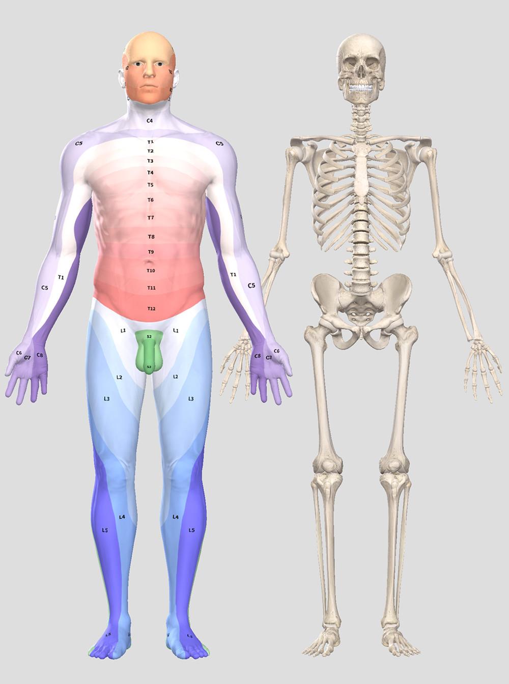
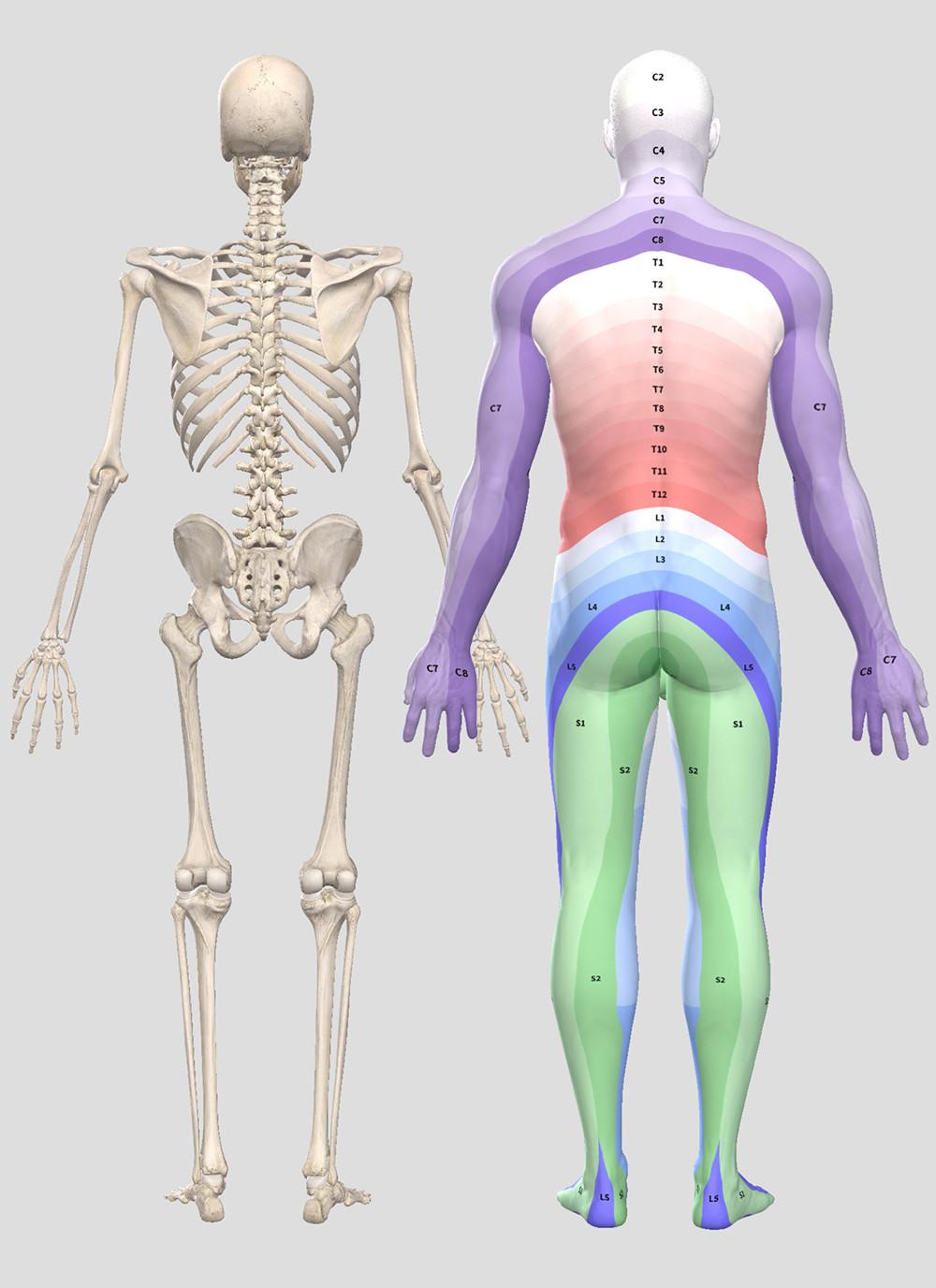
 How do you “read” your body? Three patterns, three meanings
Numbness and nerve pain can follow three basic patterns, and each means something quite different. This is the most important practical part of the article — click the tabs:
If the symptom follows a single dermatome band, the source is at the spine: most often a herniated disc is pressing on the nerve root, and less commonly narrowing caused by wear in the vertebrae, nerve inflammation after shingles or another root-related process is responsible. A typical example: when the L5 root is compressed, pain runs from the buttock along the outer thigh and lower leg towards the big toe, and lifting the foot may become weaker; with S1, pain radiates along the back of the thigh and lower leg towards the heel and little toe, and standing on tiptoe becomes weaker. An important detail clarified by medical research is that sharp, band-like radiating pain and sensory disturbance indicate root damage — whereas dull, difficult-to-localise pain that “spreads” from the lower back into the buttock and thigh is often not root-related, but joint- or muscle-related so-called referred pain.3 The two are treated differently, so the distinction matters.
Further away from the spine, the fibres of the nerve roots rearrange and join together to form peripheral nerves — a peripheral nerve therefore carries fibres from several dermatomes and supplies its own area of skin, which differs from the dermatomes. If the nerve is compressed along its course — typically in a narrow channel, a “tunnel” — the numbness follows the territory of that nerve, not the dermatome. The best-known example is carpal tunnel syndrome: the thumb, index and middle fingers become numb, typically at night — this is the territory of the median nerve, not a cervical root. This difference determines whether the neck or the wrist needs treatment. Read more: Carpal tunnel syndrome → and Treatment of tunnel syndromes →
The third pattern is neither a band nor the territory of one nerve: numbness, burning and sensory disturbance occur symmetrically on both feet (and later on the hands), in a stocking- or glove-like distribution. This is the pattern of polyneuropathy: it is not one nerve or root that is damaged, but the ends of the longest nerve fibres — most commonly because of diabetes, alcohol, vitamin B12 deficiency or chemotherapy. The distinction is vital, because in this case it is not the spine that needs investigating, but the metabolism. If this is your pattern, read on here: Causes of peripheral neuropathy and reducing pain → and Causes of burning feet syndrome →
The main dermatomes — with landmarks
You do not need to know all 31 segments; the levels below cover most symptoms. Doctors also use landmarks for orientation — you can remember some of them too:
| Root | Typical skin area / landmark | When is it considered? |
|---|---|---|
| C5 | Outer shoulder and upper arm | Cervical disc herniation, “shoulder-arm syndrome” |
| C6 | Outer forearm, thumb and index finger | One of the most common cervical herniation levels |
| C7 | Back of the arm, middle finger | Cervical herniation; the triceps reflex may be reduced |
| C8 | Inner forearm, ring and little fingers | Cervical root or ulnar nerve passing at the elbow — distinguishing between them is a medical task |
| T4 | Nipple line, in a belt-like distribution | Shingles, thoracic root pain |
| T10 | Umbilical line, in a belt-like distribution | Shingles; distinguishing internal medical pain |
| L4 | Front of the thigh, inner side of the lower leg | Lumbar herniation; the knee reflex may be reduced |
| L5 | Outer lower leg, top of the foot, big toe | The most common lumbar herniation level; weakness when lifting the foot (peroneal pattern) |
| S1 | Back of the thigh and lower leg, heel, little toe | Sciatica-like radiation; the ankle reflex may be reduced, standing on tiptoe is difficult |
| S3–S5 | “Saddle” area of the perineum and around the anus | Warning area: numbness here is an emergency sign (see below) |
A practical note: textbook dermatome maps may contradict one another because they were created using different methods, some dating back centuries — according to the evidence available today, the extent of the bands varies between individuals and neighbouring segments may overlap considerably.1,2 So if your numbness does not follow the illustration “exactly”, that does not mean you are imagining it — and conversely, a symptom that fits the map is not yet a diagnosis, but a useful clue that an examination may confirm.
 How do we use dermatomes in practice?
A herniated disc presses on the exiting nerve root, and the symptom appears in the root's dermatome and muscles: with an L5 herniation, the outer lower leg and lifting the foot are affected; with S1, the back of the leg and standing on tiptoe are affected. From the band of numbness, muscle strength and reflexes, the doctor can often predict what the MRI will show. The map helps you observe and describe the symptoms precisely: how far does the pain travel (does it go below the knee — this is an important boundary), which finger is numb, and has any movement become weaker? Read more: Herniated disc – what can you do about it? → and Sciatica symptoms and treatment →
Shingles is the most visible proof of the dermatome system. The chickenpox virus from childhood lies dormant in the nerve-root ganglia for decades, then reactivates when the body's defences are weakened — and because it awakens in a single ganglion, the blistering rash outlines exactly one dermatome: it is one-sided, belt- or band-like, with a sharp boundary at the midline. It most often occurs on the trunk (T3–L2 segments) or the face, and burning pain in the band may precede the rash by several days — at this stage, many people suspect a heart attack, gallstone or renal colic.4 Rapid recognition matters because antiviral treatment is most effective within the first 72 hours after the rash appears. Read about persistent nerve pain after shingles and complementary laser treatment: Soft laser treatment for herpes and shingles →
During spinal (epidural or subarachnoid) anaesthesia, the anaesthetist thinks in terms of dermatome levels: for obstetric epidural anaesthesia, for example, the aim is loss of sensation below T10 (the umbilical line); for a caesarean section, the block must reach T4 (the nipple line) — the level is checked on the skin using cold or pin-prick sensation. In spinal cord injury, the same system provides the language for describing the “height” of the damage (for example, “loss of sensation below the Th12 level”) and the starting point for rehabilitation. I have written a separate guide about the chances of recovery from persistent nerve damage and what to do: Nerve damage – consequences and chances of recovery →
Knowing about dermatomes also has a direct benefit at home: during TENS treatment, electrodes are typically placed over the dermatome of the painful area — the gate-control principle works most favourably when stimulation enters through the same spinal segment as the pain. With root-related radiating pain, electrodes are therefore not placed only where it hurts, but along the band: for example, with S1-type sciatic pain, one pair beside the spine on the lower back and one pair along the back of the thigh and lower leg. About programmes and settings: Pain-relief programmes – which one and what for? →
 Red flags — when numbness is an emergency
The dermatome map does not only help explain symptoms; it also helps raise the alarm. If any of the following signs are present, do not experiment with home methods. Seek medical attention or contact the out-of-hours service immediately:
- Saddle numbness + bladder or bowel problems – if the perineum, inner thighs and “saddle” area around the anus become numb, together with urinary retention or difficulty starting urination, or faecal incontinence (often accompanied by weakness in both legs): this is the picture of cauda equina syndrome — compression of the lower nerve roots in the spinal canal, which may require surgery within hours. Call an ambulance or go to emergency care immediately.
- Rapidly worsening muscle strength – if, alongside numbness, lifting the foot or gripping with the hand becomes weaker over a matter of days, or the foot “slaps” when walking: the root is losing not only sensation but also muscle function — urgent examination is needed to prevent permanent damage. About rehabilitation after paralysis: Limb paralysis – what can you do to recover? →
- Suspected shingles on the face, around the eye or the tip of the nose – a rash spreading to the tip or wing of the nose indicates involvement of the nerve supplying the eye: corneal damage may threaten vision — see an ophthalmologist or a doctor the same day, also because of antiviral treatment.4
- Bilateral, symmetrical, spreading numbness – sensory disturbance and weakness creeping upwards from both feet are not a root-related pattern: they suggest polyneuropathy or — if deterioration is rapid — Guillain–Barré syndrome, the latter being an emergency.
- Fever, weight loss, history of cancer + spinal pain – severe spinal pain that is also troubling at night and does not ease when lying down, together with these accompanying symptoms, raises suspicion of an inflammatory or tumour-related root process — this calls for investigation, not exercise or a home device.
 What does the research say? Based on readers' questions
“Why do I find several conflicting dermatome illustrations online?”
Because the maps were created in different periods using different methods — and they really do contradict one another. Most maps used today can be traced back to two families of sources: observations from the turn of the 19th and 20th centuries (rashes in patients with shingles, and surgical and injury cases), and the 1948 map by Keegan and Garrett, based on patients with herniated discs and showing long, twisted bands. Textbooks sometimes publish one, sometimes the other, and sometimes a mixture of the two.2 A systematic review from 2008 reassessed the available evidence and concluded that the classic maps were partly based on flawed studies. It produced an evidence-based version showing the bands that are most reliably present in most people, while emphasising overlap and individual variation.1 The lesson is not that the map is useless, but that it is for orientation, not a millimetre-precise diagnosis.
“If pain radiates from my lower back into my thigh, is it definitely a herniated disc?”
No — and this is one of the most common misconceptions clarified by pain research. Three different phenomena are often confused: local lower-back pain; referred pain, where dull, difficult-to-localise pain from the joints and ligaments of the spine “spreads” into the buttock and thigh (this is not nerve-root damage and typically does not extend below the knee); and true root pain, which is sharp, band-like and often like an electric shock, running along the dermatome to the lower leg and foot, and which may be accompanied by numbness and muscle weakness.3 The distinction is not pedantry: referred pain does not involve a herniation that needs surgery, and it is treated differently. If your pain radiates below the knee and follows a band, it points towards a root problem; if it stops “diffusely” in the back of the thigh, it often does not.
“Can shingles really appear only in one band?”
Typically, yes — and recognising this is the key. According to a clinical review in the New England Journal of Medicine, the typical picture of shingles is a one-sided blistering rash limited to a single dermatome that does not cross the midline. Burning pain or itching in the area may occur several days earlier; most cases affect the trunk segments.4 Involvement of two or three neighbouring dermatomes can occur, but an extensive form that does not respect band boundaries is seen mainly in patients with severely weakened immune systems — this always requires investigation. One further practical point: effective vaccination is now available for people over 50 to protect against shingles and its feared complication, postherpetic nerve pain lasting for months — ask your GP.
Frequently asked questions
It can give you a good approximation, but not a definite answer. The direction and course of the band do indicate the likely level — but because of overlap and individual variation, neighbouring roots can be confused, and the final answer comes from testing muscle strength and reflexes, and, if necessary, an MRI scan. The map's greatest benefit at home is accurate observation: if you can tell the doctor that “the pain runs from my left buttock along the outer side of my lower leg to my big toe, and I have more difficulty lifting my foot”, you can save time and avoid unnecessary tests.
They are the two sides of the same segmental system: a dermatome is the area of skin whose sensation is carried by one root, while a myotome consists of the muscles moved by that same root. This is why the two occur together: when the L5 root is compressed, the outer lower leg becomes numb (dermatome) AND lifting the foot becomes weaker (myotome). Doctors always examine them together — if only sensation is affected, it is less severe; if muscle strength is also affected, it is a more urgent sign.
Because your brain does not “see” where the pain originates; it registers which nerve pathway carries the signal. The root is compressed at the spine, but the fibres running along that root carry signals from the skin area of your leg — so the brain projects the signal to where that pathway would normally report from: the leg's dermatome. The same principle explains why a heart attack can cause pain radiating into the left arm and why a limb that has been amputated can still hurt (phantom pain).
Probably not. Root-related (dermatomal) symptoms are typically one-sided and band-like. Bilateral, symmetrical, “stocking-like” numbness is the pattern of polyneuropathy — most commonly caused by diabetes, alcohol, vitamin B12 deficiency or a thyroid problem, and its cause must be investigated with laboratory tests. The exception is an alarming situation: if bilateral numbness is accompanied by altered sensation around the perineum and bladder problems, cauda equina syndrome must be suspected — seek medical attention immediately.
A direct link between a “displaced vertebra” found by touch and numbness cannot be established by palpation: the level of root damage is estimated from the combination of sensory, muscle-strength and reflex examinations, and confirmed by imaging. Massage and manual therapy can help considerably with muscle tension and pain — but leave the diagnosis of “which vertebra” to a doctor, especially if numbness or muscle weakness is also present: these signs go beyond what can be assessed on a massage table.
 Summary – Quick overview
Sources
- Lee MWL, McPhee RW, Stringer MD. An evidence-based approach to human dermatomes. Clin Anat. 2008;21(5):363-373. DOI: 10.1002/ca.20636
- Downs MB, Laporte C. Conflicting dermatome maps: educational and clinical implications. J Orthop Sports Phys Ther. 2011;41(6):427-434. DOI: 10.2519/jospt.2011.3506
- Bogduk N. On the definitions and physiology of back pain, referred pain, and radicular pain. Pain. 2009;147(1-3):17-19. DOI: 10.1016/j.pain.2009.08.020
- Cohen JI. Clinical practice: Herpes zoster. N Engl J Med. 2013;369(3):255-263. DOI: 10.1056/NEJMcp1302674