
 What is lymphatic reconstruction surgery?
It is important to know: lymphatic reconstruction surgery is not a standalone therapy, but an option within a complex treatment package. Conservative treatment — compression garments, manual or device-assisted lymphatic drainage, exercise, and skin care — must be continued before and after surgery. The operation provides a “boost” in the condition but does not eliminate the underlying disease.
 Key point
Lymphatic reconstructive surgery today comprises four main methods: lymphovenous anastomosis (LVA), vascularized lymph node transfer (VLNT), liposuction, and debulking. Each has its own indications and they are often combined. The choice depends on stage, disease localization, and the patient’s overall condition.
The perspectives and case studies of clinicians can be read in the interview with Dr. Balázs Mohos — this article focuses on general background and professional knowledge.
Who may be a candidate for lymphatic reconstruction surgery?
The decision to operate is always individualized and based on multiple considerations. According to international guidelines and clinical practice, the most common indications are:
- Chronic lymphedema that responds to conservative therapy — patients who have received adequate complete decongestive therapy (CDT) for 6–12 months but still have persistent swelling.
- Stage II lymphedema — where fibrosis is partial and the lymphatic vessels’ structure is still amenable to microsurgical reconstruction.
- BCRL prevention and early treatment — immediate reconstruction performed during or after lymph node removal in breast cancer patients (LYMPHA, SLYMPHA techniques).
- Severe, deforming lipedema (stage 3–4) — where conservative treatment no longer provides acceptable results and tissue-volume reduction is justified for mobility and quality of life.
- Persistent, non-improving limb volume — where CDT after 6 months has not achieved at least a 30% improvement and the patient is motivated to continue postoperative routines.
Surgery is not suitable in stage III lymphostatic elephantiasis, where microsurgical reconstruction is often not feasible (lymphatic vessels have lost their functional structure) — in such cases liposuction or debulking may be considered. Active malignant disease, severe heart failure, uncontrolled hypertension, uncontrolled diabetes, and unplanned pregnancy are all contraindications.
Detailed eligibility assessment is done in a multidisciplinary team: consultation with a lymphologist, microsurgeon, physiotherapist, and oncologist.
Overview of the 4 main lymphatic reconstruction methods
The table below summarizes the four most common methods. Detailed descriptions follow in the next sections.
| Method | What it does | Who it’s for | Nature of intervention |
|---|---|---|---|
| LVA (lymphovenous anastomosis) | Microsurgical connection between lymphatic vessels and small veins — trapped fluid can be diverted directly into the venous circulation. | Stage I–II lymphedema, BCRL. | Microsurgical; local or general anesthesia; 2–4 hour operation. |
| VLNT (vascularized lymph node transfer) | Transplantation of a healthy lymph node group from another body site to the lymphedematous region with its own blood supply. | Stage II lymphedema, BCRL, postoperative limb swelling. | Microsurgical; general anesthesia; 4–6 hour operation; hospital stay. |
| Liposuction (lipedema-oriented, WAL/tumescent) | Gentle removal of subcutaneous fat while preserving lymphatic vessels. | Stage II–IV lipedema; stage III lymphedema with an adipose component. | General anesthesia; 2–4 hour operation; may require multiple sessions. |
| Debulking (Charles, Homans procedures) | Surgical removal of severely elephantiasis-affected tissue, often with skin grafting. | Stage III lymphedema where other methods are not feasible. | Surgical; general anesthesia; 3–5 hour operation; prolonged recovery. |
Clinical decisions are rarely "either-or": combined approaches are common, for example LVA + VLNT in one operation, or LVA performed months after liposuction.
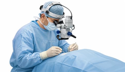
Lymphovenous anastomosis (LVA) — microsurgical connection
Lymphovenous anastomosis (or lymphaticovenous anastomosis, LVA) is the most delicate microsurgical technique. Under a supermicroscope the surgeon connects lymphatic vessels 0.3–0.8 mm in diameter to similarly sized small veins, creating a bypass-type channel. Trapped lymphatic fluid can thus flow directly into the venous blood, and lymph that would otherwise "overflow" in the classic lymph pathway can be drained.
Advantages of LVA include minimal invasiveness (often achievable through a few 2–3 cm skin incisions), typically short hospital stay (1 day), and a gentle postoperative course. Clinical results in BCRL show 60–80% improvement in limb volume in appropriately selected patient groups.
A key element of the technique is indocyanine green lymphography: dye injected under the skin fluoresces the lymphatic channels and helps the surgeon identify functioning lymphatics for anastomosis during the operation.
Postoperative phase: compression garments are usually worn from postoperative day 1–2, and the CDT routine continues. Pneumatic compression may be resumed 4–6 weeks after surgery, starting at low pressure.
Vascularized lymph node transfer (VLNT) — lymph node transplantation
VLNT differs from LVA in that it does not only create a connection but implants new, functioning lymph nodes into the affected area. The donor sites are most commonly the neck (submental), axilla, or inguinal region, from which a tissue flap containing 3–4 lymph nodes is harvested with its vascular supply and transferred to the recipient site.
The method is based on two principles: transplanted lymph nodes form their own lymphatic channels in the surrounding tissue (lymphangiogenesis), and the implanted tissue can also act as a “pump,” supporting regional lymph flow.
VLNT clinical outcomes in BCRL and lower-limb lymphedema show 50–70% improvement in limb volume and significant improvements in patient quality-of-life scores. Full effect development may take 12–18 months — regenerative processes proceed slowly.
One risk of the method is so-called "donor site morbidity" — secondary lymphedema may develop at the donor site. Modern techniques (e.g., omentum flap) minimize this risk, but it remains an important consideration in patient counseling.
Liposuction for lipedema and lymphedema
Lipedema- and lymphedema-oriented liposuction differs from classic cosmetic liposuction by using techniques optimized to preserve lymphatic vessels — most commonly water-assisted liposuction (WAL) or tumescent techniques. The targeted tissue removal addresses:
- Stage II–IV lipedema: the pathological adipose tissue that constitutes the bulk of limb volume.
- Stage III lymphedema with an adipose component: fat accumulated during the chronic process that conservative therapy no longer mobilizes.
Liposuction for lipedema is therapeutic, not cosmetic: clinical studies with 5–10 year follow-up show pain reduction by 70–90%, limb volume reduction by 30–50%, and significant improvement in mobility. Two-thirds of patients report dramatic improvement in quality of life.
In severe cases multiple surgical sessions are required, as a surgeon usually recommends removing a maximum of 4–5 liters of fat per session to maintain fluid balance. The full program may involve 2–4 sessions spaced 3–6 months apart.
Postoperative phase: 4–6 weeks of intensive compression garment wear; pneumatic compression may be resumed after 4–6 weeks (with medical approval). For detailed clinical background on lipedema see the Lipedema (fat edema) — symptoms and treatment article.
Debulking surgeries — for severe elephantiasis
Debulking (volume-reducing) procedures — Charles procedure, Homans procedure, Thompson technique — are used in stage III lymphostatic elephantiasis when the limb is so deformed that microsurgical reconstruction is no longer feasible. The operation removes excess skin and subcutaneous tissue with extended resection and often covers the surface with skin grafts.
This method is the most burdensome for patients, requiring longer hospital stays and recovery times (4–8 weeks). Results can be dramatic in limb size and function, but cosmetic outcomes are often suboptimal (visible scarring, graft areas). This method is now less commonly first-line — LVA + VLNT or combinations with liposuction often achieve similar volume reduction in a gentler way.
Debulking is primarily considered when the patient is resistant to all conservative treatments, has recurrent erysipelas complications, and limb size severely limits mobility.
 Expected outcomes and clinical evidence
The evidence base for lymphatic reconstructive surgery has strengthened significantly in the last decade. One of the highest-level reviews was published by Donahue and colleagues in 2023:
Donahue et al. (2023) — review on BCRL prevention and treatment
Modern BCRL prevention and treatment rely on a multimodal approach: sentinel lymph node removal as standard care, early detection using tape measurement and bioimpedance, and complete decongestive therapy (CDT) as the fundamental intervention. Among surgical methods, lymphovenous anastomosis (LVA), vascularized lymph node transfer (VLNT), and immediate lymph node reconstruction (LYMPHA, SLYMPHA) are gaining prominence. Low-level laser therapy (LLLT) and advanced pneumatic compression devices (APCD) are promising adjuncts.1
It is realistic to set expectations regarding outcomes:
- Early-stage LVA: 60–80% volume reduction in appropriately selected patients, with 50–70% symptom improvement.
- VLNT in stage II: 50–70% volume reduction and significant improvement in quality-of-life (LYMQOL) scores.
- Lipedema liposuction: 30–50% volume reduction, 70–90% pain reduction, with long-term (5–10 year) durable results if conservative therapy continues.
- Debulking in stage III: dramatic volume reduction, but often suboptimal cosmetic outcome.
Full effect development takes time: LVA typically 3–6 months, VLNT 12–18 months, liposuction 6–12 months. Patients should be informed of these timelines before surgery to set realistic expectations.
Postoperative care and long-term management
The postoperative phase after lymphatic reconstruction surgery is as important as the procedure itself. The following points apply to all methods:
1. Daily wear of compression garments. Intensive compression class (II–III) is recommended for the first 4–6 weeks after surgery, followed by long-term maintenance compression. After liposuction compression is particularly important — the limb’s final shape is "set" during the first 6 weeks.
2. Pneumatic compression. From 4–6 weeks postoperatively, with the treating physician's agreement, starting at low pressure. Detailed device selection logic can be found in the Lymphatic massage device category.
3. Manual lymphatic drainage (MLD). Performed by a trained lymph therapist 1–3 times weekly during the first 3 months after surgery, then less frequently. Method details are in the Manual lymphatic drainage guide.
4. Exercise therapy. Controlled exercise in compression garments (swimming, walking, cycling) from the first 4–6 weeks, with gradually increasing intensity.
5. Skin care and infection prevention. The skin of the affected limb is particularly vulnerable — continue daily moisturizing after wound healing and promptly treat any injury or insect bite.
6. Long-term specialist follow-up. Follow-up every 3 months in the first year, then annually. Volume monitoring is done with tape measurement or bioimpedance.
Deeper guides in the cluster
Lymphatic reconstructive surgery is a specialized area within the compression-therapy ecosystem. Full cluster guides:
- Lymphatic reconstruction surgery — interview with Dr. Balázs Mohos — specialist opinion and practical experience
- Lymphedema — types, causes and stages — guide
- Managing lymphedema at home — conservative treatment protocol
- Manual lymphatic drainage — manual and device-assisted lymphatic massage — basis of postoperative routine
- Lipedema (fat edema) — symptoms and treatment — lipedema-focused guide
- Radiation therapy and lymphedema — BCRL context
- Lymphatic massage device — multi-indication hub — pneumatic compression device selection
 When is lymphatic reconstruction surgery NOT recommended?
The surgical decision is made by a multidisciplinary team, and the following conditions significantly reduce or exclude the possibility of intervention:
Absolute and relative contraindications
- Active malignant tumor in the treated region — oncologic consultation and tumor-free status are required before surgery.
- Severe heart failure — the strain of long surgery and general anesthesia may be excessive.
- Uncontrolled hypertension or diabetes — postoperative wound healing risk is significantly increased.
- Active skin infection (erysipelas, cellulitis) — intervention cannot be performed until the infection heals.
- Severe peripheral arterial disease — individual assessment; surgical risk.
- Unsuitable patient cooperation — if postoperative routines (compression, exercise, follow-up) cannot be guaranteed, results will not be durable.
- Stage III lymphostatic elephantiasis (for LVA, VLNT) — lymphatic vessels have lost functional structure; liposuction or debulking may be considered here.
 Important information
Lymphatic reconstruction surgery does not replace conservative treatment — CDT must be continued before and after surgery. Surgical decisions are always made by a multidisciplinary team (lymphologist, microsurgeon, physiotherapist). The experience of specialists and institutional background available in Hungary and abroad are decisive for outcomes.
Frequently asked questions
No. Lymphatic reconstruction surgery can substantially improve symptoms and reduce limb volume, but it does not eliminate the nature of the disease. Postoperative use of compression garments, exercise, and pneumatic compression must continue — this is the key to durable results. Clinical studies with 5–10 year follow-up show that postoperative results are lasting if postoperative routines are maintained.
LVA (lymphovenous anastomosis) is most effective in stage I–II lymphedema where lymphatic vessel structure is still preserved. VLNT (vascularized lymph node transfer) is often used in stage II or as a complement to LVA, particularly in BCRL or lower-limb lymphedema. The exact choice is determined by the surgeon based on imaging studies (indocyanine green lymphography).
After LVA there is usually a 1–2 day hospital stay, then 2–4 weeks of limited activity. Full effect develops over 3–6 months. For VLNT the hospital stay is typically 3–5 days, 4–6 weeks of limited activity, and full effect develops over 12–18 months. Return to office work is generally possible after 2–4 weeks; heavy physical work after 6–8 weeks.
Yes, LVA, VLNT and lipedema-oriented liposuction are available at larger surgical centers. The number of Hungarian specialists with surgical experience and international training is steadily growing. For clinical decision-making it is worth consulting several specialists and considering their institutional background, case experience, and clinical results. The specialist perspective is available in the interview with Dr. Balázs Mohos.
After LVA and VLNT, pneumatic compression is generally resumed 4–6 weeks after surgery, starting at low pressure as agreed with the treating physician. After liposuction it is also 4–6 weeks, but compression garments are especially important from day 1 postoperatively. After debulking surgery it is often 6–8 weeks due to longer wound healing. The exact protocol is coordinated by the surgeon and physiotherapist.
The funding situation is complex: for certain indications (e.g., reconstruction after BCRL) public insurance may partially or fully cover the procedure; in other cases (e.g., lipedema liposuction) the patient may bear the cost. Exact information should be obtained from the chosen specialist and institution. In private care prices vary significantly by institution and method.
 Summary — Lymphatic reconstructive surgery in brief
Sources
- Donahue PMC, MacKenzie A, Filipovic A, Koelmeyer L (2023). Advances in the prevention and treatment of breast cancer-related lymphedema. Breast Cancer Research and Treatment. DOI: 10.1007/s10549-023-06947-7
- Su L, Huang H, Tong Y, and colleagues (2025). Intermittent pneumatic compression devices for the prevention and treatment of breast cancer-related lymphedema – a systematic review and meta-analysis. Supportive Care in Cancer. DOI: 10.1007/s00520-025-10159-8