What Can Cause a Shadow on the Lung? The Chest X‑Ray Examination
If you see a doctor or go to emergency care for chest pain, trauma or shortness of breath, a chest X‑ray is usually taken (often along with other tests such as an ECG). X‑ray is an imaging method that is typically one of the first diagnostic tests used to clarify diseases affecting the chest. I wrote about why it is done and what can be seen on it.

A chest X‑ray can show the heart, the lungs, the central part of the thoracic cavity (the mediastinum — the space between the two lungs), the large blood vessels, the airways, as well as the ribs and spinal bones. X‑rays can also reveal fluid accumulated in or around the lungs or air surrounding the lung (pneumothorax).
By analyzing the details of the image, the doctor can more easily determine whether there is a structural abnormality of the heart, a collapsed lung, pneumonia, broken ribs, emphysema, a tumor or any other similar disease or condition. It is important that tumorous changes, whether benign or malignant, are detected in time through thorough examinations and screenings.
In some diseases, several chest X‑rays may be taken a few days apart to monitor whether the problem has improved or worsened.
What is a chest X‑ray?
A chest X‑ray is a test that helps assess the condition of the lungs and other organs in the chest. The examination uses X‑rays that pass through the body and create an image of the organs and tissues. This test is particularly useful in diagnosing lung cancer, pneumonia, heart disease and other chest conditions.
During the exam, the patient is usually asked to stand or lie down and hold their chest still while the X‑rays pass through the body. The procedure takes only a few minutes and is completely painless. The images obtained are interpreted by a radiologist who identifies abnormalities and diseases.
This test is especially important for early detection of lung cancer and other chest diseases, because early diagnosis is key to effective treatment. Chest X‑rays are typically performed in the following situations:
- Suspicion of lung cancer
- Suspicion of pneumonia
- Suspected heart disease
- Chest pain or shortness of breath
- Follow‑up of previous chest diseases
In addition to a chest X‑ray, other imaging tests such as CT or PET/CT may be performed to help achieve a more precise diagnosis. These tests provide more detailed images of the chest organs and assist in establishing an accurate diagnosis and planning appropriate treatment.
Why is it done?
The chest X‑ray is a common type of test and is often among the first investigations performed when your doctor suspects heart or lung disease. It is most frequently used when pneumonia is suspected. It can also help check how you are responding to a given treatment.
The image can reveal many things inside your body, including:
The condition of the lungs. X‑rays can detect tumors, infections, or air trapped around the lung that can cause lung collapse. Timely diagnosis of tumorous changes is particularly important for effective treatment. Chronic lung diseases such as emphysema or cystic fibrosis, and complications related to these conditions, can also be identified. Patchy shadows on the image can suggest pneumonia and show the extent of the inflammation.
Lung problems related to the heart. The image can reveal changes or problems in the lungs that result from heart disease. For example, fluid in the lungs can be a consequence of congestive heart failure.
The size and outline of the heart. Changes in heart size and shape can indicate heart failure, fluid around the heart, or valve problems.
Blood vessels. Because the outlines of large vessels near the heart—the aorta and the pulmonary arteries and veins—are visible on X‑rays, they can reveal aortic aneurysms (marked dilation of the main artery, often with thinning of the vessel wall), other vascular problems or congenital heart disease.
Calcified deposits. X‑rays can show calcification in the heart or vessels, which can damage heart valves, coronary arteries, the heart muscle, or the pericardium (the protective sac around the heart). Calcified nodules in the lungs most often remain as remnants of an old, healed infection.
Fractures. Rib fractures, spinal fractures or other bone problems may also be seen on the image.
Postoperative changes. Useful for monitoring recovery after chest surgery (for example heart, lung or esophageal surgery). The position of tubes placed during surgery, any air leaks, and areas of fluid or air accumulation can be checked.
Pacemaker, defibrillator or catheters. Leads of pacemakers and defibrillators are attached to the heart to help regulate heart rate and rhythm. Catheters are small tubes used for internal drug delivery or dialysis. After placement of such medical devices, a chest X‑ray is usually taken to ensure everything is in the correct position.
Risks of a chest X‑ray
You may wonder, especially if you have repeated X‑rays, how much radiation your body is exposed to.
Research on modern X‑ray equipment shows that the amount of radiation from these images is low—lower than the exposure you receive from natural environmental sources of radiation (for example cosmic rays from space).
The latest X‑ray machines operate with minimal doses of ionizing radiation, so the exposure to the body remains minimal and the test is considered safe. Overall, however, I recommend not requesting a new examination every week—accept only those tests that are truly necessary.
Although the benefits of X‑rays clearly outweigh the risks, in some cases a gonad shield may be used during imaging, especially if multiple images are required. X‑rays can damage genetic material, so when women and men are actively trying to conceive, the reproductive organs and the lower abdomen should be shielded from unnecessary radiation. Therefore, inform the technician if you are pregnant or might be pregnant. Men should also inform the examiner if they are currently trying to conceive.
How to prepare
Before a chest X‑ray, you will usually need to expose your upper body (remove clothing from the waist up). Jewelry should also be removed from the upper body, as clothing and jewelry can obscure X‑ray images or cover certain areas and may lead to incorrect results.
What to expect
During the exam you will be positioned between the X‑ray machine that produces the rays and a detector that records the image digitally (or, with older machines, on X‑ray film). The X‑rays pass through your body, including bone, and the detector captures the resulting “imprint.”
You may need to adopt different body positions so that both the front and the side details of the chest can be seen. Part of the lung is difficult to assess with X‑ray because overlapping internal organs (such as the diaphragm, heart, and large vessels) can make interpretation harder. Areas behind the ribs, the lung apices, and the thoracic inlet are often obscured on conventional X‑ray film.
For the frontal view you will stand close to the plate, raise or press your arms to your sides, and roll your shoulders forward. The radiographer will ask you to take a deep breath and hold it for a few seconds. Holding the breath helps keep the chest and lungs still so the heart and lungs appear clearer on the image.
For lateral (side) views one shoulder is pressed to the plate and the arm is raised above the head. You may again be asked to take a deep breath and hold it.
The X‑ray itself is usually painless. You will not feel anything when the radiation passes through your body.
If you have difficulty standing due to your physical condition, the exam can sometimes be performed while you are sitting or lying down.
Results
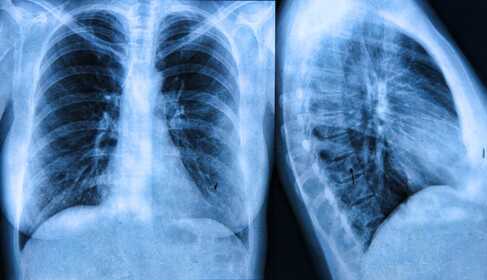
A chest X‑ray produces a black‑and‑white image showing the organs in the chest. Structures that block the rays appear white; structures that allow the rays to pass through appear dark.
Bones appear white because they are dense. Your heart also appears as a lighter area. Your lungs are filled with air and block very little radiation, so they appear darker on the images.
The interpretation of images produced by X‑rays (and other imaging tests) is performed by a radiologist (an imaging specialist). When analyzing the films, they look for clues that may indicate heart failure, fluid around the heart, tumor, pneumonia, fracture, injury or other abnormal conditions. It is important that tumorous changes, benign or malignant, are identified through careful examinations and screenings.
Today, many lung tumors are still discovered incidentally on chest X‑rays taken for screening or other reasons, often because other, more advanced screening programs are not widespread.
It is appropriate that, after a thorough expert evaluation of the chest X‑ray, your doctor discusses the result with you, including what treatments or further tests or procedures may be needed.
Common abnormalities
When a shadow is detected on a chest X‑ray, it can indicate several different abnormalities. Such shadows can be harmless and of no risk in some cases, while in others further investigations are necessary.
These shadows are often reported as a rounded shadow or a solitary nodule on the report. These are most often benign lesions, such as residual scarring from a past inflammation, a calcified lymph node, a developmental abnormality, or a benign tumor. These findings typically do not require intervention, but regular (X‑ray) monitoring is recommended.
The size and shape of the shadow provide important information. Small nodules under about 4 mm rarely indicate malignancy, whereas larger nodules over about 8 mm or those with irregular shapes require further tests.
The growth rate of a nodule is also informative; if it does not change in size over a long period, it is probably benign.
Smoking, exposure to asbestos, or a family history of lung cancer can increase the risk of a malignant lesion. In such cases regular screening and medical follow‑up are particularly important.
If a shadow is seen on an image, the reporting physician will look for your previous films (if any). They compare the current image with earlier ones to determine whether it was present before and whether it has changed. If necessary, additional imaging tests such as CT or PET‑CT may be performed, and in some cases a tissue sample (biopsy) may be indicated to establish the exact diagnosis.