
 What is this spot? – The skin "rusting"
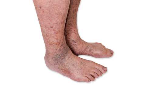
The discoloration is actually caused by “rust”, i.e. deposited oxidized iron in the skin. It forms when red blood cells leak out of the smallest vessels (capillaries) in larger numbers into the tissues. When these red blood cells break down, the iron released from their hemoglobin converts into hemosiderin. It is stored in the subcutaneous tissue and appears on the shin as a brownish-reddish spot.
The spot itself is not painful and does not itch – but from a medical perspective it is an important sign: it indicates persistent venous circulation disturbance and, in advanced cases, may be a precursor of venous leg ulcer.
 Key point
The reddish-brown spot is "only" a symptom, but it can indicate three important underlying causes: chronic venous insufficiency (CEAP C4), post-thrombotic syndrome or a precursor of venous leg ulcer. Hemosiderin deposited in the skin cannot be removed, but by treating the underlying circulatory disorder (exercise, compression, home devices) further deterioration can be prevented.
 How does hemosiderin deposition develop?
Your heart pumps blood through the arteries to the rest of your body; then it returns to your heart through the veins. Circulation is maintained by your heart, the diaphragm and respiratory muscles, and by contraction of your muscles during movement.
Veins contain "valves" — so-called venous valves — that permit blood to flow only one way (back toward the heart). As long as these valves close properly, they prevent blood from falling back toward the leg.
The problem arises when these valves weaken or the vein dilates. If this happens, the valve "lets go" and upward blood flow worsens. More and more blood remains in the veins; they swell, dilate and become tortuous (commonly called varicose veins).
If the condition persists, the increasingly distended vein wall becomes overstretched and begins to "leak" — red blood cells escape into the surrounding tissues. From the hemoglobin of the destroyed red blood cells iron is released, converted to hemosiderin, and deposited in the skin, creating the characteristic reddish-brown spot.
What lies behind the spot? – Venous hypertension and venous insufficiency
Reddish-brown discoloration of the lower leg is most often caused by venous problems, especially when pressure in the leg veins is persistently elevated. This condition is known as venous hypertension or venous insufficiency.
Your risk is higher if:
- your job requires long periods of standing or sitting without movement;
- your parents or siblings have similar leg problems (family tendency);
- you are being treated for varicose veins or leg ulcers;
- you have had deep vein thrombosis in the past — details in the thrombosis article, PTS section.
The classic, often years-unnoticed background is chronic venous insufficiency: if at the end of a long day your leg feels heavy ("as if you were wearing lead boots"), hurts or swells, that is not a good sign. The appearance of brownish-red spots and thin, dry skin around them indicates CEAP C4 stage — detailed classification in the chronic venous insufficiency article.
What can the spot indicate? – Differential diagnosis
The reddish-brown spot can point to three main backgrounds:
| Background | Characteristics | Where to turn? |
|---|---|---|
| Chronic venous insufficiency (CEAP C4) | Varicose disease, tortuous-dilated veins, evening ankle and calf swelling | Chronic venous insufficiency article |
| Post-thrombotic syndrome (PTS) | Gradually developing swelling after previous deep vein thrombosis, hemosiderin spot | Thrombosis article, PTS section |
| Lipodermatosclerosis / ulcer precursor | Skin is hard, inflamed, thickened; an indentation appears or a slowly healing wound | Venous leg ulcer (EMS protocol) or soft-laser protocol |
If leg edema is also present, the triage article helps with differential diagnosis: Leg swelling (edematous leg) causes and treatment.
What to do if reddish-brown spots appear?
If the spots have appeared, you are somewhat late for prevention — nevertheless, see a vascular surgeon clinic and request an evaluation. It is necessary to determine the severity of the problem; based on that, treatment can be set up to prevent further worsening.
At the clinic, in addition to history and physical examination, a Doppler ultrasound is usually performed. Its essence is to determine blood flow velocity in the leg vessels using ultrasound and to compare both sides.
Further tests may be needed to assess diabetes or cardiac involvement.
How can the spot and its cause be treated?
Hemosiderin deposited in the skin cannot be removed — the discoloration will therefore accompany you for life. What you can do is prevent or slow further deterioration and treat the underlying venous insufficiency.
Skin hydration
The skin of the discolored area thins, becomes dry and may crack. Avoid drying soaps. Use a moisturizing cleanser or cosmetic. Bath water should not be too hot — that also dries the skin. Do not use coarse towels or bath mitts, you may injure the thin skin.
Compression therapy
There are two types of compression options: elastic compression stockings made of stretchable material or machine pneumatic massage (intermittent pneumatic compression), often referred to as a lymphatic massage machine. Compression therapy supports venous blood flow, effectively reduces swelling, promotes healing and helps prevent wounds or ulcers.
Functional electrical stimulation (FES)
A significantly cheaper (though somewhat less effective) method than pneumatic compression is electrical stimulation of the lower limb muscles. The muscle contractions triggered by the impulses support limb circulation similarly to muscle movement. Favorable results can be expected after a few weeks of daily 20–30 minute sessions.
Exercise – the natural “pump”
The natural motor of venous circulation is regular muscle contraction, i.e. movement. To support venous circulation, brisk walking, jogging or cycling are most suitable. Move as often as possible: ideally 20–30 minutes once or twice a day, or a single 40–50 minute session. In this case, more is better!
 Home devices – physiotherapy
If active movement is limited (reduced mobility, joint problems, severe varicose disease), home medical devices can support the calf muscle pump effect.
Pneumatic compression (lymphatic massage machine)
Power Q-2200 lymphatic massage machine
A mid-range device with multiple treatment programs. Recommended for venous insufficiency, CEAP C4 stage, hemosiderin spots and post-thrombotic follow-up.
Power Q-1000 Plus lymphatic massage machine
Entry-level home device for mild-to-moderate venous complaints.
Power Q-1000 Premium lymphatic massage machine
Advanced home device with multiple programs and greater comfort – for advanced venous insufficiency.
Muscle stimulators (EMS / FES) devices
Myolito TENS/EMS/FES device
Multifunctional electrotherapy device with TENS, EMS and FES programs – for functional electrical stimulation to support the calf muscle pump.
Rehalito EMS muscle stimulator
Simple, affordable 2-channel device – designed specifically for rehabilitation and circulation support.
Elite SII TENS/EMS device
Multifunctional device with 100 programs: EMS and pain-relieving TENS functions.
 Before you start treatment – contraindications
For safe use, know the contraindications.
When NOT to use the lymphatic massage machine (IPC)?
- Acute deep vein thrombosis
- Severe, decompensated heart failure
- Acute skin infection, open wound on the treatment area, without treating physician's permission
- Active malignant tumor in the treatment area, without treating physician's permission
When NOT to use the muscle stimulator (EMS / FES)?
- Implanted pacemaker or defibrillator
- Suspected acute thrombosis
- Infected or inflamed skin in the treatment area
- Malignant tumor in the treatment area
- Pregnancy (abdomen and low back area)
 Important information
If you suffer from any heart disease or circulatory problem, consult your treating physician before starting treatment. Read the device user manual.
 Prevention – what you can do to avoid it
Since shin discoloration is a consequence of venous insufficiency, if you don't want reddish-brown spots on your legs, prevent them. The most effective method is daily regular exercise. Start in childhood and never stop exercising.
My advice – a simple daily routine
- 30–40 minutes of brisk walking (can be split into two sessions);
- stand up every hour during sedentary work and mobilize the calf (rise onto toes and lower to heels 15–20 repetitions);
- do not sit with your legs crossed;
- avoid prolonged exposure to hot water for the feet – long hot foot baths, saunas or thermal baths are unfavorable;
- elevate your legs at the end of the day for 15–20 minutes.
 Scientific background
Pathophysiology of hemosiderin deposition in chronic venous insufficiency
The review by Eberhardt and Raffetto (2014) on the pathophysiology of chronic venous insufficiency describes in detail the mechanism of hemosiderin deposition: venous hypertension → capillary extravasation → red blood cell destruction → hemosiderin release → tissue deposition.1
Compression therapy in venous insufficiency
According to the consensus recommendation by Rabe et al. (2022), compression of 20–30 mmHg is advised from CEAP C4; graduated compression stockings reduce edema and progression of hemosiderin deposition by supporting venous return.2
Hemodynamic effect of pneumatic compression
In the clinical study by Kakkos et al. (2001), intermittent pneumatic compression favorably influenced venous circulation and alleviated symptoms of chronic venous insufficiency.3
ESVS 2022 European clinical guidelines
De Maeseneer et al. edited the European Society for Vascular Surgery (ESVS) 2022 clinical practice guidelines for the management of chronic venous disease of the lower limbs, which represent the current standard for CEAP classification and treatment algorithms.4
Frequently asked questions
Hemosiderin deposited in the skin cannot be removed — the spot usually remains for life. What you can do: by treating the underlying venous insufficiency you prevent further spread of the spot and the development of more serious complications (lipodermatosclerosis, leg ulcer).
A hemosiderin spot is a specific skin sign (iron deposition) that can develop on several backgrounds: chronic venous insufficiency (CEAP C4), post-thrombotic syndrome (PTS) or lipodermatosclerosis. PTS is an overarching clinical syndrome following prior deep vein thrombosis — the hemosiderin spot is only one of its signs. Details in the thrombosis article, PTS section.
If the skin around the hemosiderin spot becomes hard, thickened, inflamed (lipodermatosclerosis), or if the area shows thinned, fissured skin, this is a direct precursor to venous leg ulcer (CEAP C5–C6). In such cases consult a vascular surgeon promptly. Detailed home treatment protocols: venous leg ulcer with EMS or soft-laser treatment.
Yes, in chronic venous insufficiency (from CEAP C3) pneumatic compression can favorably influence symptoms. It is contraindicated in suspected acute thrombosis, and in the presence of active skin infection or open wound it may be used only with the treating physician's permission. See section 8 for details.
 Summary – quick overview
Sources
- Eberhardt RT, Raffetto JD (2014). Chronic venous insufficiency. Circulation. PubMed: 24834437
- Rabe E et al. (2022). Risk factors for chronic venous disease and compression therapy in chronic venous disease: international consensus. Phlebology. PMC7874878
- Kakkos SK et al. (2001). Improved hemodynamic effectiveness of a new intermittent pneumatic compression system in patients with chronic venous insufficiency. J Vasc Surg. PubMed: 11700495
- De Maeseneer MG et al. (2022). European Society for Vascular Surgery (ESVS) 2022 Clinical Practice Guidelines on the Management of Chronic Venous Disease of the Lower Limbs. Eur J Vasc Endovasc Surg. PubMed: 35027279