
 Why is the distinction important?
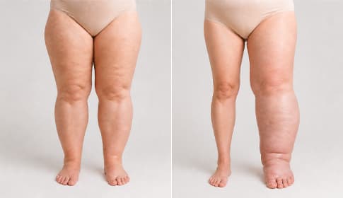
The aim of this article is to provide a practical guide to distinguishing the two conditions — with visual signs, clinical tests and a comparison of treatment strategies. A definitive diagnosis should always be made by a specialist (lymphologist, vascular surgeon), but the information below will help you get oriented and be prepared for consultations.
 Key point
Lipedema is a disorder of adipose tissue — treatment mainly targets the tissue mass and inflammation. Lymphedema is a fluid-circulation disorder — treatment primarily targets fluid drainage. The two conditions can co-occur; this is called lipolymphedema.
 Anatomical-pathological basics — what happens in the tissues?
The two diseases occur in different tissue layers and cause swelling by different mechanisms.
In lipedema subcutaneous fat accumulates pathologically and symmetrically. The causes are complex: polygenic genetic predisposition, hormonal factors (estrogen sensitivity), and chronic low-grade inflammation. The increased fat mass is painful, tender and bruises easily. The lymphatic system is initially intact — it only becomes secondarily involved due to mechanical tissue pressure in stages 3–4.
In lymphedema the capacity of the lymphatic system is reduced or obstructed. This may be congenital (primary lymphedema — rare, developmental disorder) or secondary to external events (surgery, radiotherapy, injury, infection, chronic venous congestion). The movement of protein-rich interstitial fluid is impaired, increasing local inflammation and eventually leading to connective tissue scarring (fibrosis). The skin remains intact, but the tissue gradually hardens.
Because of these two fundamental differences, treatment also differs:
- In lipedema the main goals are: control of the pathological adipose tissue + reduction of inflammation + stabilization of quality of life.
- In lymphedema the main goals are: fluid drainage + slowing tissue changes + infection prevention.
Detailed clinical background can be read in the Lipedema (fat edema) symptoms and treatment and the Lymphedema — forms, causes and stages guides.
Visual and symptomatic differentiation
The detailed table below summarizes the eight main differences used in clinical practice. A person in good health can even go through it themselves:
| Feature | Lipedema | Lymphedema |
|---|---|---|
| Distribution | Symmetric, bilateral (hips, thighs, calves, upper arms) | Typically unilateral (except congenital primary forms) |
| Foot / back of hand | Not involved, remains slender — “cuff sign” | Usually involved, swollen |
| Stemmer sign | Negative (the skin fold can be pinched up) | Positive (the skin fold cannot be pinched up) |
| Skin sensitivity | Tender, bruises easily | Initially normal, later tight and indurated |
| Pitting (indentable swelling) | Absent or minimal | Yes, typical in early stages |
| Pain | Typical, also on pressure | Initially minimal, later chronic |
| Sex distribution | Almost exclusively women | Both sexes (depending on indication) |
| Typical triggering event | Hormonal changes (puberty, pregnancy, menopause) | Surgery, radiotherapy, infection (secondary); congenital (primary) |
The main message of the table: the two conditions have clear clinical features that in most cases give guidance even before specialist examination.
 Clinical tests and investigations
Four main clinical examination methods are used for accurate differential diagnosis. Some of these can be done at home, others require consultation with a specialist.
1. Stemmer sign (can be done at home)
The classic clinical test for diagnosing lymphedema. Try this: at the base of the second toe attempt to pinch the skin into a fold with your fingers. If it lifts easily into a fold (negative Stemmer sign) → you probably have lipedema. If the skin CANNOT be lifted (positive Stemmer sign) → you likely have lymphedema. The test is not infallible, but in clinical practice it is a strong initial indicator.
2. Pitting test (can be done at home)
Press the edematous area with your fingers for 5–10 seconds, then remove them. If a visible indentation remains that returns over minutes → pitting-positive (typical of stage 1 lymphedema). If there is no visible indentation → likely lipedema or more advanced (above stage 2) lymphedema. This test detects the presence of fluid — lipedema is primarily increased tissue mass, not fluid, so pitting is absent or minimal.
3. Tape-measure limb measurement (home or specialist)
Measuring limb circumference at the same points weekly with a tape measure helps assess trends (direction of change). In lipedema the two sides are roughly symmetric (both thighs similar size); in lymphedema (especially secondary/BCRL) the affected side is markedly larger. A difference greater than 2 cm is an early sign.
4. Bioimpedance spectroscopy (BIS) — specialist
A modern, sensitive method that measures extracellular and intracellular fluid volumes. It is very useful for early detection of BCRL (before symptoms!). It may be available in clinics of clinical lymphedema therapists and lymphology specialists.
5. Lymphoscintigraphy and MR lymphangiography — hospital specialist
Imaging techniques used for definitive diagnosis in more complex cases (e.g., congenital primary lymphedema, lipolymphedema). They accurately show lymphatic vessel function and anatomy. These examinations are performed at specialist hospitals.
Lipolymphedema — when both conditions are present
Lipolymphedema is the clinical situation when a lipedema patient develops secondary lymphedema in stages 3–4. The typical course is:
- stage 1 lipedema develops (smooth skin, tender tissue),
- over years or decades it progresses to stages 2–3 (mattress-like skin, coarse irregularity),
- the increased tissue mass mechanically compresses (compresses) lymphatic vessels and lymph nodes,
- lymphatic capacity decreases and soft, fluid-like swelling appears on the already enlarged limb,
- finally classic lipedema signs (bilateral, free foot) and lymphedema signs (foot involvement, positive Stemmer sign) appear together.
Treatment of lipolymphedema combines the strategies for both core conditions: elements of complete decongestive therapy (CDT) (compression garment + manual or mechanical lymphatic drainage + skincare + exercise) are primary. Pneumatic compression is recommended at low pressure (30–40 mmHg) under medical supervision in this case. Surgical options (lipedema-oriented liposuction + sometimes LVA or VLNT) may also be considered — details in the Lymphatic reconstruction surgery guide.
Stage-based treatment strategies for lipedema are detailed in the Lipedema stages 1–4 cluster article.
What does the distinction mean for treatment?
Diagnostic accuracy also determines the treatment strategy. The table below summarizes the most important differences:
| Treatment element | Lipedema | Lymphedema |
|---|---|---|
| Goal | Control tissue mass, reduce inflammation, relieve pain | Fluid drainage, slow tissue change, prevent infection |
| Compression garment | Class II (23–32 mmHg) — daily wear | Class II–III (23–46 mmHg) — daily wear, individually fitted |
| Pneumatic compression pressure | 30–60 mmHg | 30–50 mmHg |
| Exercise | Emphasized — under compression: swimming, walking, cycling | Emphasized — muscle pump function under compression |
| Diet | Anti-inflammatory approach (Mediterranean, ketogenic) | Moderate sodium intake, hydration, weight control |
| Special treatments | In severe cases liposuction (stages 3–4) | In severe cases microsurgery (LVA, VLNT) |
| Manual lymphatic drainage (MLD) | Useful adjunct | Primary — one of the main pillars of CDT |
 Device selection by indication
The choice of pneumatic compression device differs by stage and indication. The most important difference is the pressure range:
- Lipedema stages 1–2: 4-chamber home device at 30–60 mmHg. Power Q-1000 Plus entry-level, Power Q-2200 value-for-money, Power Q-1000 Premium advanced.
- Lipedema stage 3: 6-chamber professional Power Q-8060 with a finer sequential pattern.
- Lipedema stage 4 / lipolymphedema: 12-chamber top professional Power Q-8120, at low pressure, under medical supervision.
- Lymphedema stages 1–2: 4-chamber home device at 30–50 mmHg. Q-1000 Plus, Q-2200 or Q-1000 Premium.
- Lymphedema stage 3: 6- or 12-chamber professional device (Q-8060 or Q-8120) with finer patterning.
- BCRL (post-breast-cancer arm swelling) prevention: 4-chamber IPC ≤40 mmHg, >2 weeks. Q-1000 Plus or Q-2200 are ideal.
The full selection logic is in the Lymphatic massage device — what it is for, how to choose? guide, and the multi-indication hub is in the Lymphatic massage device category.
When should you see a specialist?
Accurate diagnosis — whether lipedema or lymphedema — is always the responsibility of a specialist (lymphologist, vascular surgeon, plastic surgeon with lipedema practice). Consultation is especially important in the following situations:
- Persistent unexplained swelling: whether symmetric (suspect lipedema) or unilateral (suspect lymphedema), persistent swelling warrants specialist consultation.
- New swelling after oncological surgery: arm or leg swelling that appears after breast, cervical or prostate cancer treatment — even years later — should be assessed by a lymphologist.
- Unclear clinical picture: if home self-tests (Stemmer sign, pitting) gave uncertain results or the symptoms do not clearly fit either picture.
- Recurrent skin infection (erysipelas, cellulitis): both conditions commonly complicate with infections — repeated episodes require specialist protocols.
- Considering surgical options: consider liposuction or microsurgery (LVA, VLNT) for stage 3–4 lipedema or severe lymphedema. Details in the Lymphatic reconstruction surgery guide.
A specialist medical perspective in clinical practice can be read in the interview with Dr. Balázs Mohos in this interview.
Clinical evidence in treatment of the two conditions
The evidence base for the two conditions has developed differently over the past decades. The studies below support the creation of indication-specific protocols.
Donahue et al. (2023) — BCRL prevention and treatment review
Modern BCRL care is based on a multi-pillar approach: sentinel lymph node removal, early detection with tape measurement and bioimpedance, complete decongestive therapy (CDT), pneumatic compression, microsurgical techniques. The review summarizes the key evidence for stage-based lymphedema treatment protocols.1
Su et al. (2025) — BCRL meta-analysis, 1397 patients
Combined results of 14 randomized clinical trials: pneumatic compression significantly reduces the incidence of breast cancer–related lymphedema (RR=0.36; 95% CI 0.22–0.58). The optimal protocol: ≤40 mmHg, >2 weeks, ≤24 months after surgery. This is lymphedema-specific evidence support.2
Atan and Bahar-Özdemir (2020) — Lipedema RCT, revised Wold criteria
In 33 women with severe (stage 3) lipedema, the combination of complete decongestive therapy (CDT) + exercise produced the largest reductions in limb volume, pain and physical function. IPC + exercise also showed significant improvement compared with the control group. This provides lipedema-specific evidence focusing on combined measures of pain and volume.3
Herbst et al. (2025) — APCD lipedema RCT
Thirty days of home APCD use significantly reduced leg volume, extracellular and intracellular fluid, and subcutaneous adipose tissue thickness as confirmed by ultrasound. In the lipedema population this study clearly supports a multimodal approach — differing from lymphedema evidence by measuring SAT (subcutaneous adipose tissue) change as well.4
The evidence base for treating the two conditions thus has different foci: in lymphedema fluid volume and prevention are central, while in lipedema tissue volume, pain and quality of life are the focus. This explains why accurate diagnosis is necessary to select the treatment protocol.
Deeper guides in the cluster
Guides for the two pillars:
- Lipedema (fat edema) symptoms and treatment — pillar guide
- Lipedema stages 1–4 — stage-based guide
- Lipedema — fat edema category — stage-based product recommendations
- Lymphedema — forms, causes and stages — pillar guide
- Treating lymphedema at home — conservative treatment protocol
- Lymphatic drainage — manual and mechanical lymphatic massage — physical methods
- Lymphatic reconstruction surgery — surgical options
- Radiotherapy and lymphedema — BCRL context
- Lymphatic massage device — multi-indication hub — device selection
- Lymphatic massage device — what it is for, how to choose? — technical guide
 What should you watch for if you have lipedema or lymphedema?
There are several conditions in which you should definitely consult a doctor before using home treatments for either condition. These are general contraindications for pneumatic compression and compression garments.
Contraindications
- Acute deep vein thrombosis or suspected DVT — treatment only with medical permission and monitoring.
- Severe heart failure — increased venous return may cause decompensation.
- Active skin infection (erysipelas, cellulitis) — not recommended until infection heals; may be restarted after completion of antibiotic treatment.
- Severe peripheral arterial disease — individual assessment and low pressure indicated.
- Untreated high blood pressure — start after stabilization and medical approval.
- Active malignant tumor in the treated region — only with oncologist approval.
 Important to know
An accurate diagnosis and treatment plan with your treating physician or lymphologist is essential. Home IPC and compression garments should always be used only as adjuncts to medical and physiotherapeutic treatment, and specialist consultation is required for new symptoms, increasing swelling, pain or skin changes.
Frequently asked questions
Yes, this clinical situation is called lipolymphedema and usually develops in stage 3–4 lipedema. The increased tissue mass mechanically compresses the lymphatic system, leading to secondary lymphedema. Treatment then combines protocols for both core conditions and often proceeds under specialist supervision.
Preliminarily yes — home Stemmer sign and pitting tests give a good initial guide. If the Stemmer sign is negative and swelling is symmetric + the foot is free → likely lipedema. If the Stemmer sign is positive and swelling is unilateral + the foot is involved → likely lymphedema. However, a definitive clinical diagnosis should be made by a specialist (lymphologist, vascular surgeon).
Yes. Power Q series central units are multi-indication — the same compressor can be used for lipedema and lymphedema. The difference is in the settings: pressure, treatment time and program. Always agree on an indication-specific protocol with your treating physician or lymphatic therapist. Power Q-1000 Premium and higher models are suitable for both indications.
Yes, it is part of the basic treatment for both conditions. In lipedema Class II compression (23–32 mmHg) is typical and recommended for daily wear. Compression reduces pain, improves venous return, and slows stage progression. In lymphedema compression garments are even more emphasized (Class II–III) because they stabilize the mobilized fluid.
The Stemmer sign is a classic clinical test for diagnosing lymphedema. At the base of the second toe try to pinch the skin into a fold with your fingers. If the skin lifts easily into a fold (negative Stemmer sign), lymphedema is unlikely (or in an early stage). If the skin CANNOT be lifted into a fold (positive Stemmer sign), lymphedema is probable. The test is simple and can be done at home, but a positive result requires specialist consultation.
Unfortunately, neither is completely reversible. Lipedema is a chronic adipose tissue disorder and cannot currently be completely eliminated — but it can be well controlled with multimodal treatment, and in severe cases liposuction can reduce the stage. Lymphedema is also a chronic lifelong condition, but complete decongestive therapy and modern microsurgical techniques (LVA, VLNT) can achieve significant improvement. Early detection and consistent treatment are key for both conditions.
 Summary — Lipedema or lymphedema?
Sources
- Donahue PMC, MacKenzie A, Filipovic A, Koelmeyer L (2023). Advances in the prevention and treatment of breast cancer-related lymphedema. Breast Cancer Research and Treatment. DOI: 10.1007/s10549-023-06947-7
- Su L, Huang H, Tong Y, and colleagues (2025). Intermittent pneumatic compression devices for the prevention and treatment of breast cancer-related lymphedema – a systematic review and meta-analysis. Supportive Care in Cancer. DOI: 10.1007/s00520-025-10159-8
- Atan T, Bahar-Özdemir Y (2020). The Effects of Complete Decongestive Therapy or Intermittent Pneumatic Compression Therapy or Exercise Only in the Treatment of Severe Lipedema: A Randomized Controlled Trial. Lymphatic Research and Biology. DOI: 10.1089/lrb.2020.0019
- Herbst KL, Zelaya C, Sommerville M, Zimmerman T, McHutchison L (2025). An Advanced Pneumatic Compression Therapy System Improves Leg Volume and Fluid, Adipose Tissue Thickness, Symptoms, and Quality of Life and Reduces Risk of Lymphedema in Women with Lipedema. Life (Basel). DOI: 10.3390/life15050725